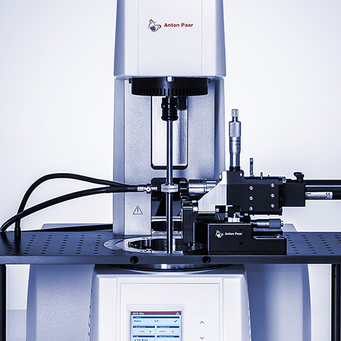
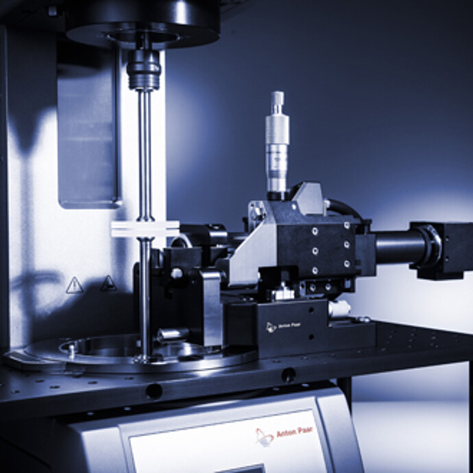
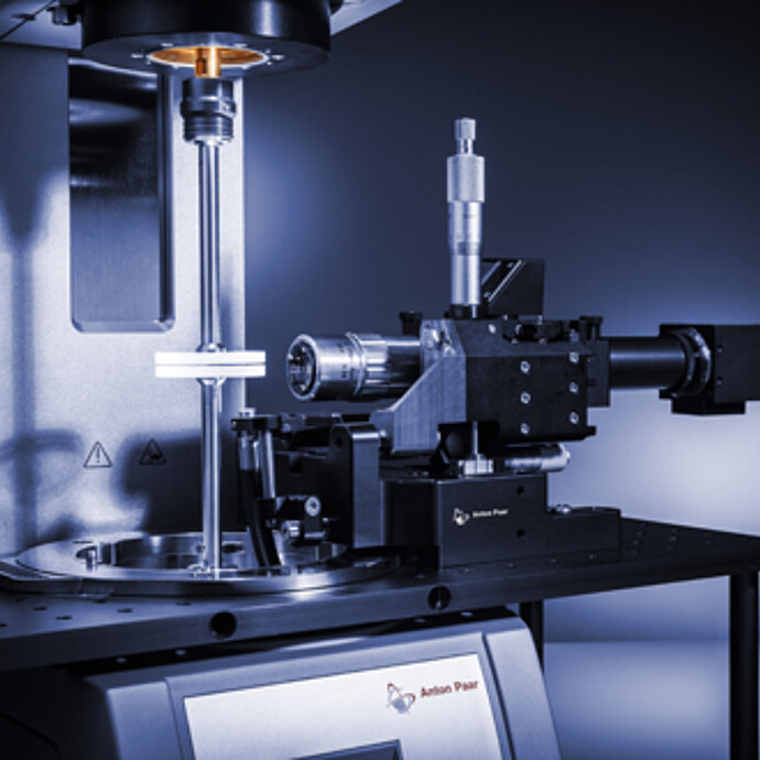
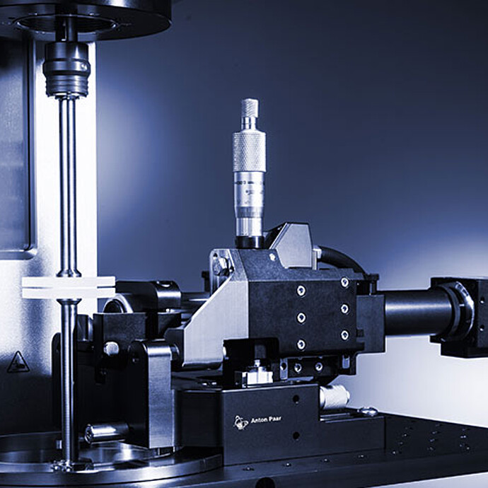
Option for a Rheometer with two EC Drives:
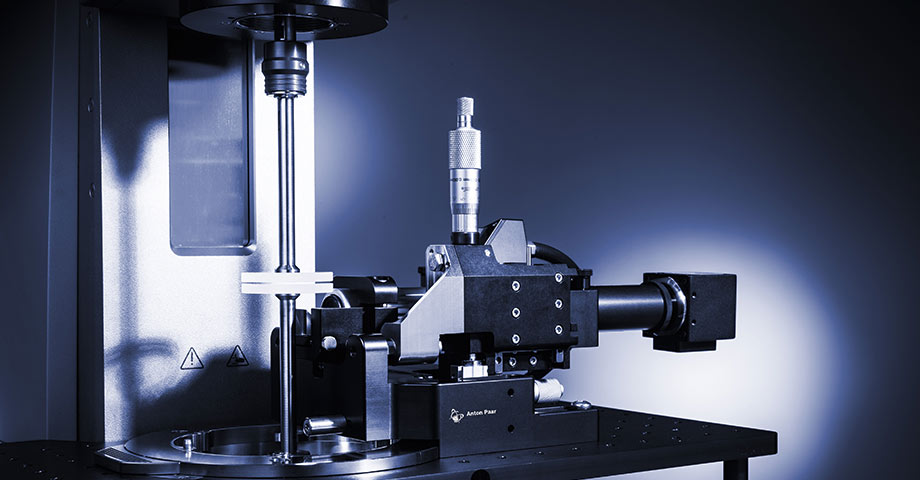
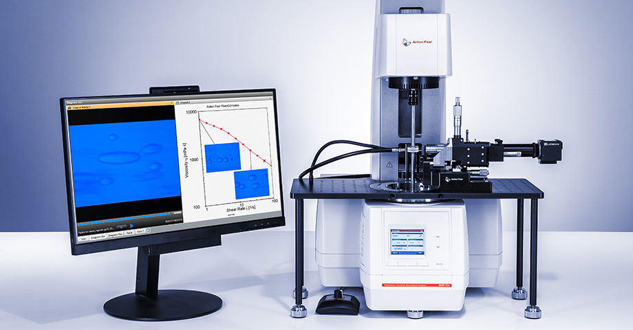
Rheo-Microscopy Setup
- Flow visualization in the stagnation plane
- Speed distribution between the two drives manually adjustable
- Direct assignment of images and videos to rheological data
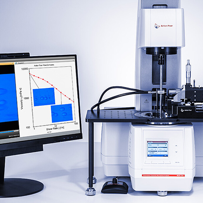
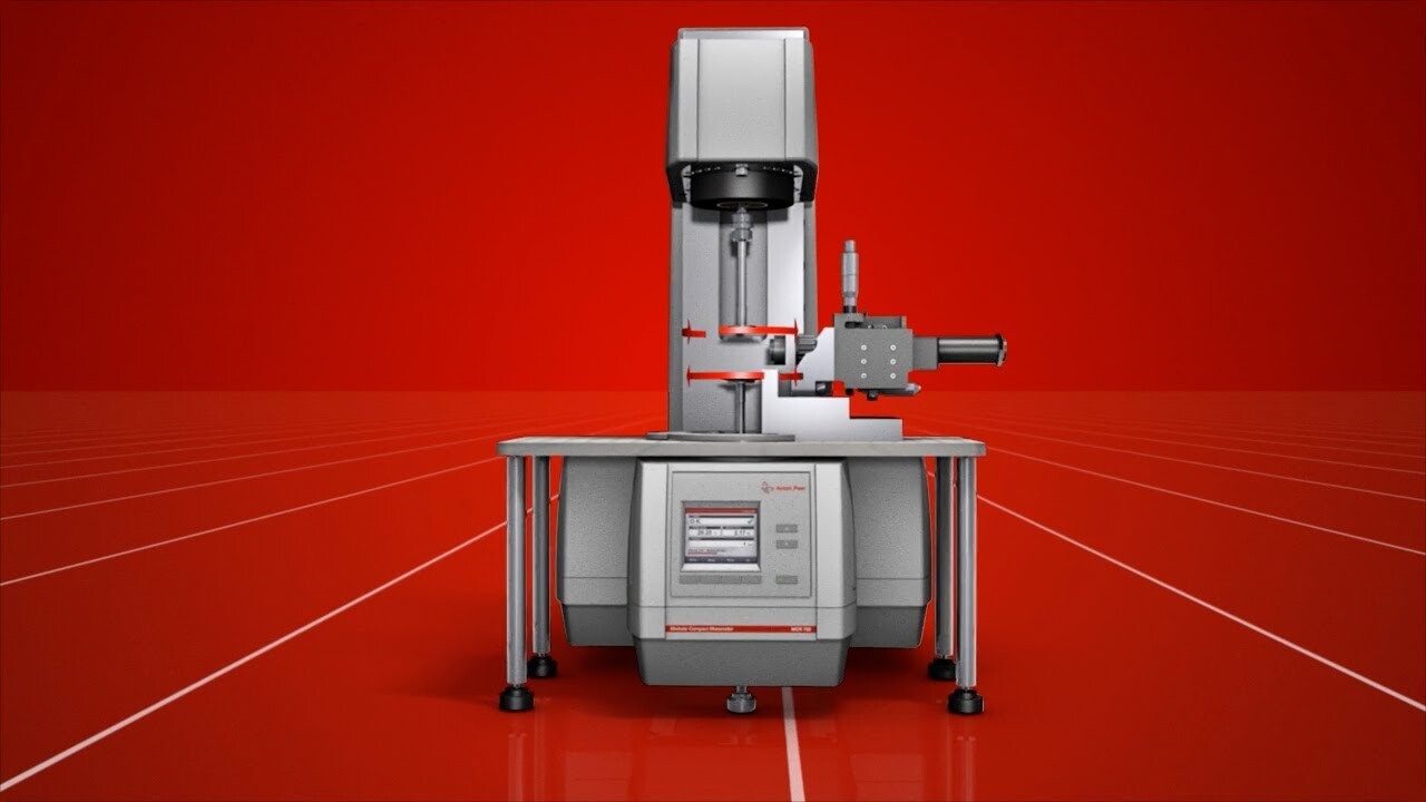
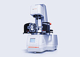
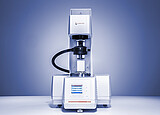
Rheo-microscopy is an established technology for visualizing the inner structures of samples while applying shear and deformation forces. The rheo-microscopy setup for a configuration with two EC-drives is a system to perform measurements in counter-rotation and counter-oscillation mode, producing a stagnation plane where the sheared structure can be kept in the microscope’s field of view while accurate rheological data is determined. In this way, breakage of agglomerates, alignment, deformation, and relaxation processes can be visualized for individual structures.
Key features
Complete flow visualization in the stagnation plane
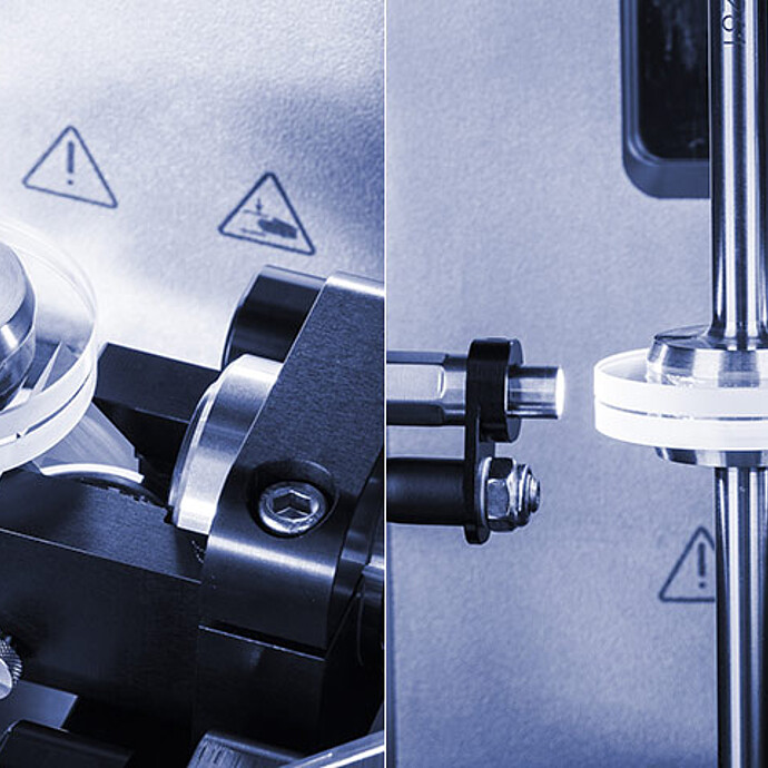
The rheometer’s counter-rotation and counter-oscillation modes can be used to produce a stagnation plane in which the observed structure is sheared but remains in a fixed position. The stagnation plane constantly keeps the focused structure in the microscope’s field of view while accurate rheological data is obtained.
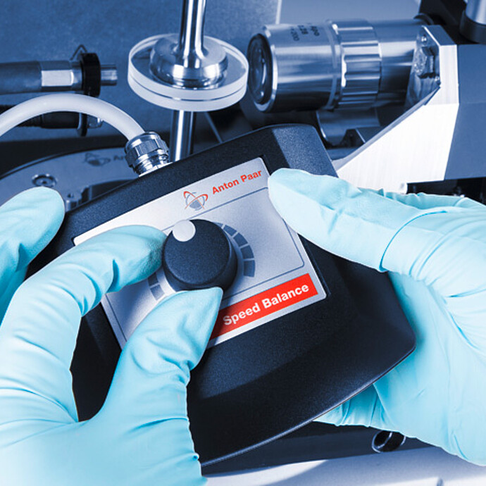
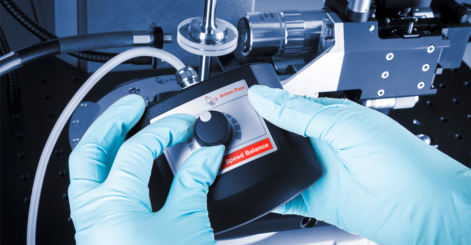
Speed balance allows you to trap and fix the structures
Using the speed balance in counter-rotation mode allows you to change the speed distribution between the two drive units and move the stagnation plane vertically through the sample. In this way, any structure of interest can be kept in the field of view and individually investigated and evaluated without changing the shear rate applied to the sample.
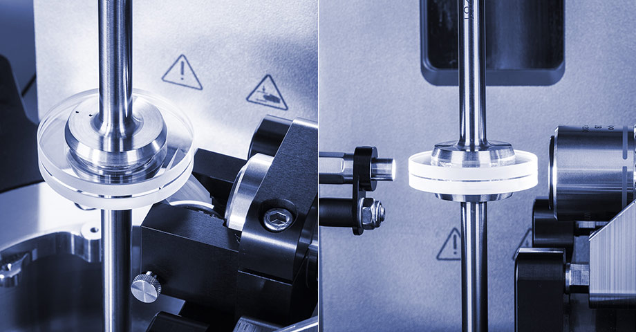
Flexible type of access and illumination
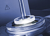
Besides the possibility of sample focusing through the lower plate via a mirror, the system can also be positioned to focus into the sample gap from the side. The holder can be moved in the x- and z-direction for sample scanning in radial and axial direction. The sample is illuminated by a light guide, either along the optical axis or over a suitable light guide holder.
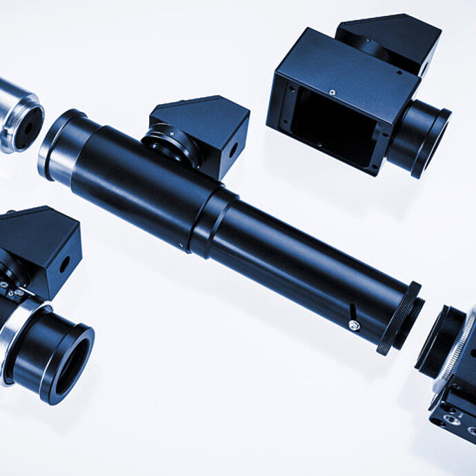
Options for polarized, non-polarized, and fluorescence microscopy
The modularity of the Anton Paar microscope setup allows you to simply switch between different optical techniques. When polarized light or fluorescence is required, the corresponding optical module can be easily mounted while all the other components remain unchanged.
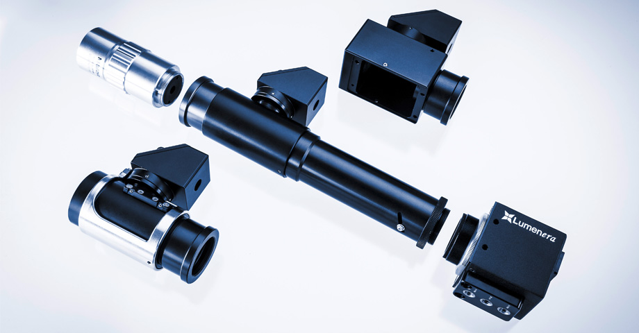
Image recording and display
The rheometer software controls both the rheometer and CCD camera which enables automated image and video recording during the measurement. Furthermore, the software offers a direct assignment of images and videos to rheological data to highlight correlations between macroscopical and structural properties directly.
Anton Paar Certified Service
- More than 350 manufacturer-certified technical experts worldwide
- Qualified support in your local language
- Protection for your investment throughout its lifecycle
- 3-year warranty
Documents
-
TwinDrive Microscopy as an Advanced Rheometric Tool for Structure Investigations Application Reports
